Uncontrolled blood sugar levels. Prior standard of care wound treatment was ineffective.
Wound Presentation:
Partial thickness wound on the medial aspect of the left hallux with irregular borders.
Prior Treatment:
The patient had previously been treated at various times with collagenase enzymatic debridement gel, collagen powder, calcium algenate, silver algenate, medical honey, hydrogel, and triple antibiotic ointment alongside of serial debridement and wound cleansing.
Study Methodology:
Use of Lavior’s Inula-based wound product on a daily basis.
Product(s) Used in Study:
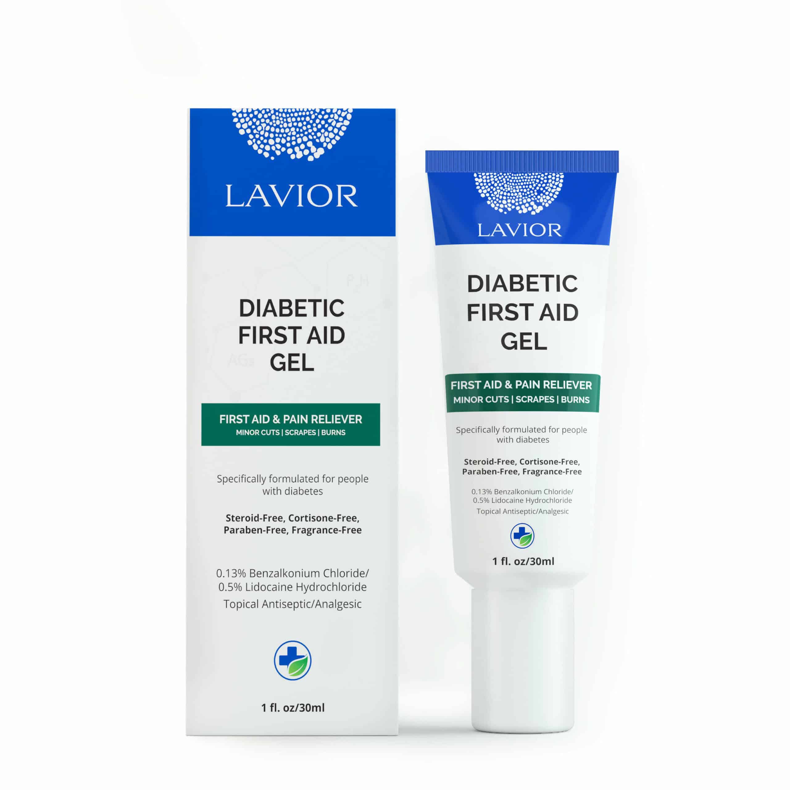
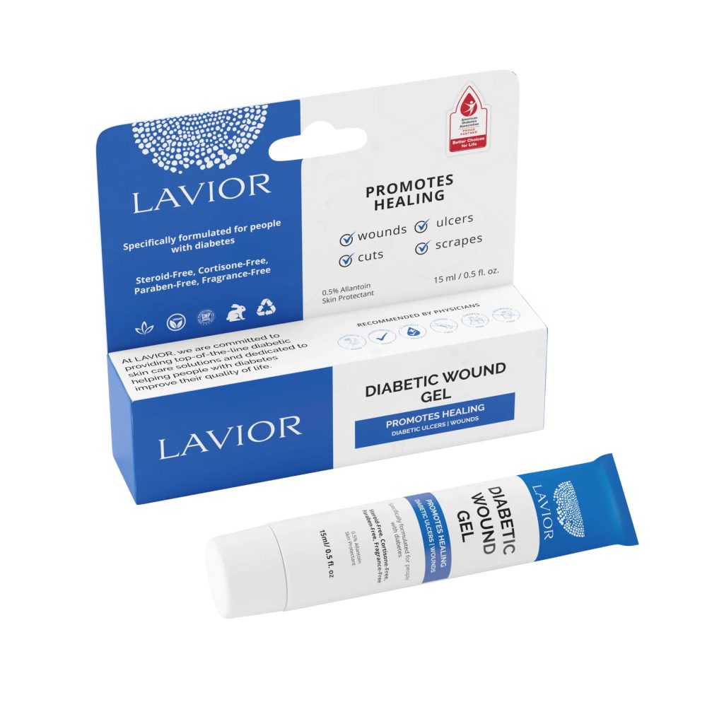
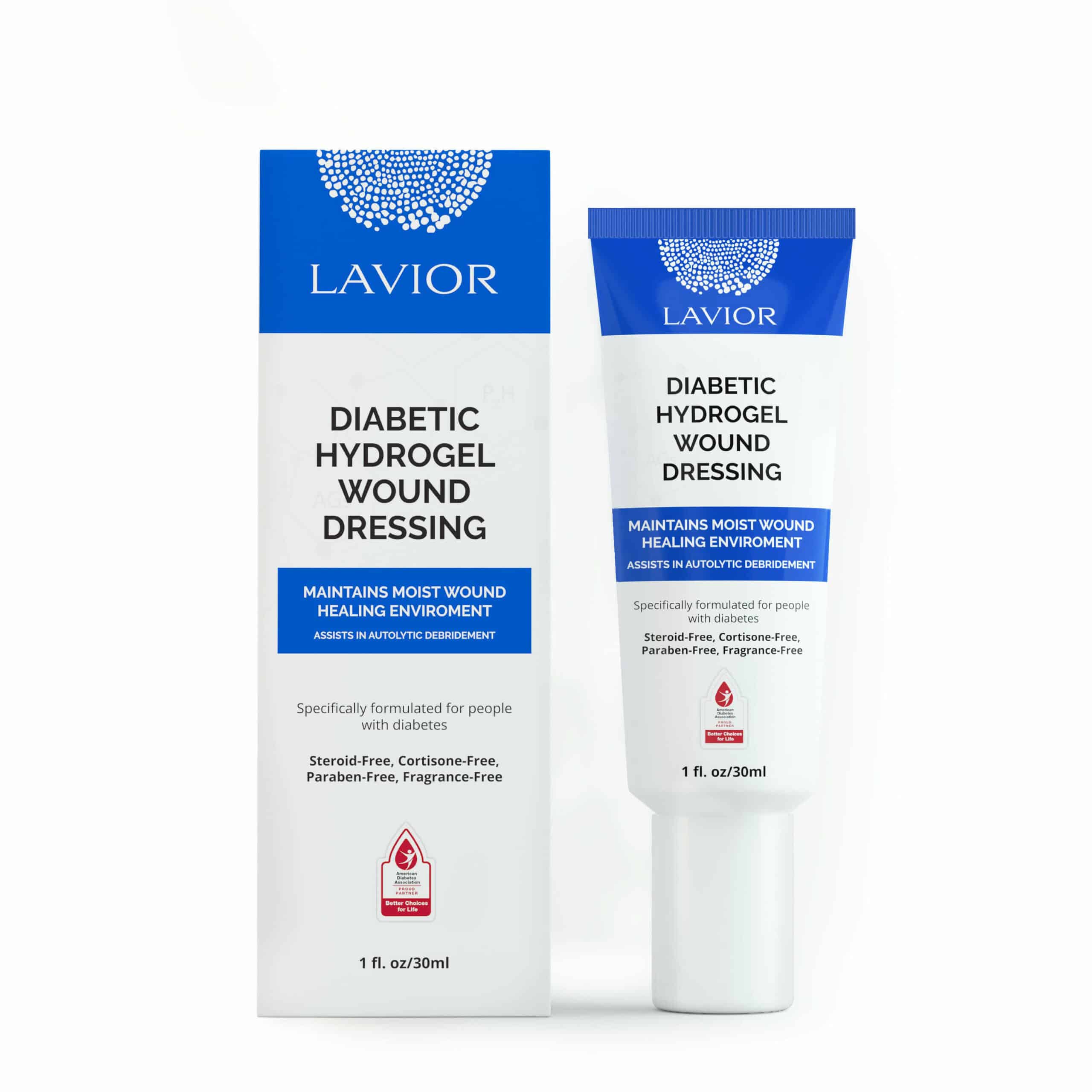
D-Care Diabetic Wound Care.
Study Duration:
4 Weeks – 4 Week Predictive Marker Study.
Treatment Narrative
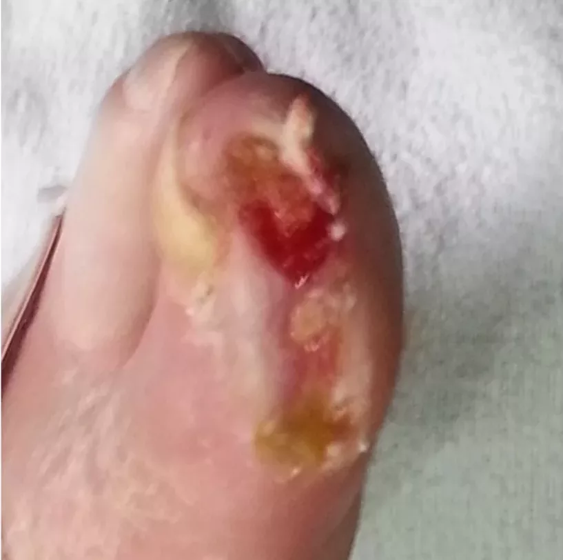
INITIAL PRESENTATION
Wound size: 4.5 cm. Stage 2 wound. Fibrotic tissue is evident with unhealthy granulation tissue. Presence of biofilm due to the chronicity of the wound.
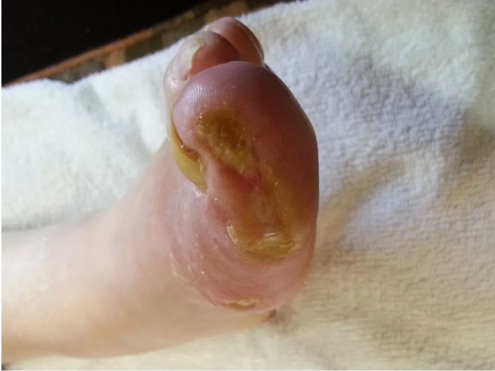
AFTER 1 WEEK OF TREATMENT
The wound is decreasing in size and getting shallower. The wound edges are taking on a drier appearance. The inflammation is decreasing, and the next phase of healing has begun.
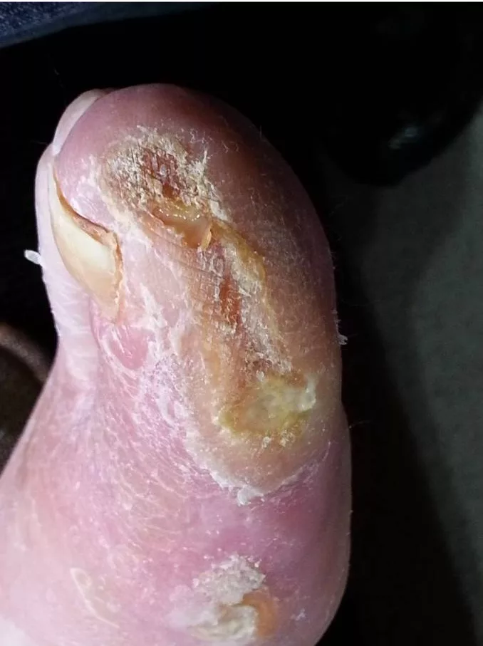
AFTER 2 WEEKS OF TREATMENT
The biofilm has been disrupted and is now eradicated. Eschar is now forming, and the wound continues to get smaller. The wound edges are approximating end to end.
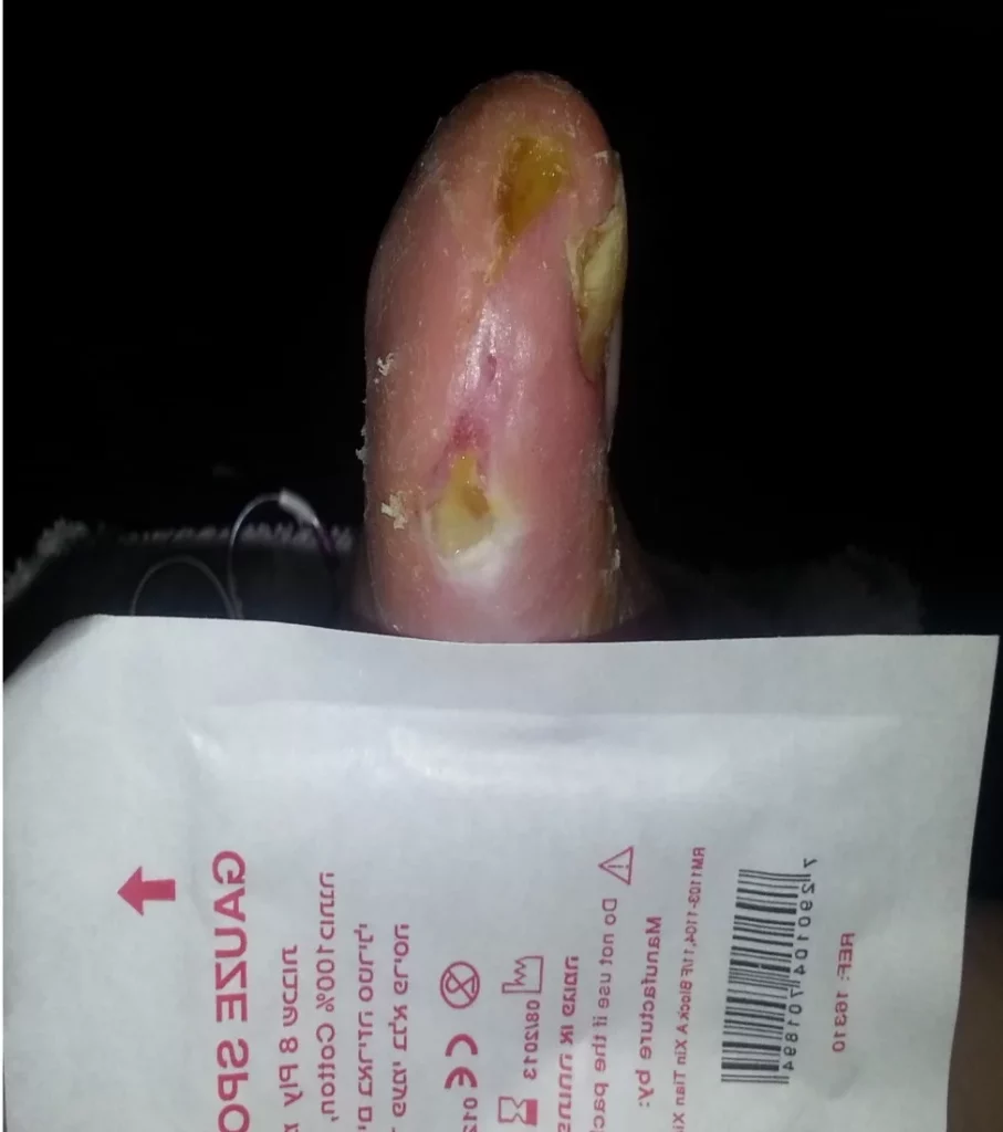
AFTER 4 WEEKS OF TREATMENT
Wound size: 0.75 cm. The vast majority of the wound has closed, and epithelial tissue is visible. The most proximal portion of the wound is still open but very shallow.
RESULT SUMMARY
During this 4-week period, wound closure is at 83%. As evident from the photos, all fibrotic tissue and biofilm were converted to granulation and new epithelial tissue.