Wounds heal in four overlapping stages: hemostasis, inflammation, proliferation and remodeling. Each stage has a specific job, and a problem at any point, most often during inflammation, can slow healing or stall it altogether.
Knowing the stages makes it easier to understand what a wound should look like as it heals and when something may be off.
What are the 4 stages of wound healing?
The stages run in order but overlap, and their timing depends on the size and depth of the wound and on your overall health.
- Hemostasis (minutes to hours): blood vessels narrow and platelets form a clot to stop the bleeding. The clot also acts as a temporary scaffold for repair.
- Inflammation (roughly the first several days): white blood cells move in to clear bacteria, debris and damaged tissue. Some redness, warmth and swelling around a fresh wound is normal during this stage.
- Proliferation (from a few days to several weeks): new tissue, called granulation tissue, fills the wound, small blood vessels regrow, and skin cells migrate across the surface to close it.
- Remodeling (weeks to a year or more): collagen is reorganized and strengthened. The scar gradually flattens and fades, although repaired skin never regains quite the strength of the original.
Why do some wounds get stuck in inflammation?
A wound can stall when inflammation does not switch off on schedule. Instead of moving on to building new tissue, it stays in a cycle of breakdown and repair.
Common reasons include:
- Bacteria or dead tissue in the wound: the immune system keeps responding as long as debris or bacteria, sometimes organized as a biofilm, remain.
- Poor blood flow: without enough oxygen and nutrients, cells cannot move to the next stage.
- High blood sugar: diabetes can disrupt immune cell function and the signals that end inflammation.
- A wound bed that is too dry or too wet: both can interfere with normal cell activity.
- Repeated pressure or friction: ongoing trauma keeps reopening the injury.
People with diabetes, circulation problems or aging skin are especially likely to have wounds that stall. When that happens for weeks, the wound may become chronic. Learn the warning signs in What Makes a Wound Chronic?
How can you tell which stage a wound is in?
You cannot always tell exactly, but visible changes offer clues. Early on, a wound may be red, a little swollen and tender. As it moves into proliferation, you may see pink or red, slightly bumpy tissue and a wound that is getting smaller. During remodeling, the wound is closed and the scar slowly changes color and texture.
For a checklist of good signs and red flags, see How to Tell if a Wound Is Healing Properly. If you are recovering from a procedure, What Does Normal Wound Healing Look Like After Surgery? covers incisions specifically.
How can you support each stage?
You cannot rush the stages, but you can remove obstacles that slow them down:
- Keep the wound clean: rinse gently and change dressings as directed.
- Keep it moist, not soaked: moist wound healing is widely considered more favorable than letting a wound dry out and form a hard scab.
- Protect it: cover the wound and avoid pressure or rubbing on the area.
- Support your body: eat enough protein, stay hydrated, avoid smoking and, if you have diabetes, keep blood sugar in your target range.
- Get help early: a wound that is not improving after about two weeks, or shows signs of infection, should be checked by a healthcare provider.
How Lavior's Multipurpose Wound Gel fits in
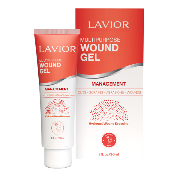
Lavior™ Multipurpose Wound Gel helps maintain a moist wound environment when used for the indications described on its label. Follow the product instructions and your healthcare professional's wound care plan. Like every Lavior formula, it is built around Inula AGS-RIED, Lavior's proprietary extract of the Mediterranean plant Inula viscosa.
Sources
- Eming SA, Martin P, Tomic-Canic M. Wound repair and regeneration: mechanisms, signaling, and translation. Sci Transl Med. 2014;6(265):265sr6. doi:10.1126/scitranslmed.3009337
- Guo S, DiPietro LA. Factors affecting wound healing. J Dent Res. 2010;89(3):219–229. doi:10.1177/0022034509359125